Picture Of Forearm Muscles And Tendons : Superficial Anterior Forearm Muscle Anatomy And Function Kenhub / Long flexor tendons extend from the forearm muscles through the wrist and attach to the small bones of the fingers and thumb.
Picture Of Forearm Muscles And Tendons : Superficial Anterior Forearm Muscle Anatomy And Function Kenhub / Long flexor tendons extend from the forearm muscles through the wrist and attach to the small bones of the fingers and thumb.. Originates from the anterior surface of the ulna and attaches to the. Most of these originate from the lateral epicondyle. The superficial group arises mostly from the posterior aspect of the lateral epicondyle of the humerus by a common tendon. It turns… inflamed common flexor tendon cft. The pronator teres has two heads of.
They receive additional fibers from the deep fascia of the forearm near the elbow, and from the septa which pass from this fascia between the individual muscles. A square shaped muscle found deep to the tendons of the fdp and fpl. The anterior forearm muscles are divided into 3 muscular layers ; The superficial anterior forearm muscles share a common origin on the common flexor tendon that arises from the medial epicondyle of humerus. A common muscle belly is shared by all the fingers.
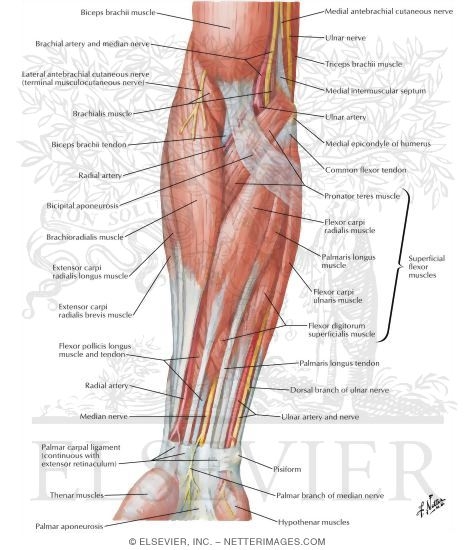
Muscles Of Forearm Superficial Layer Anterior View from www.netterimages.com While the ventral side of the forearm is not exactly less complicated than the dorsal side, it appears less complicated on the. Find stockbilleder af forearm muscles tendons i hd og millionvis af andre royaltyfri stockbilleder, illustrationer og vektorer i shutterstocks samling. The longer the muscles in the forearm are (and therefore the shorter their tendons are), the easier it will be to develop them. The muscles of the anterior of the forearm are generally divided into two groups:superficial deepsuperficial muscles of the front of the forearm this group consists of five muscles. Also, pollicis means thumb in latin. It is separated from the anterior compartment by the interosseous membrane between the radius and ulna. 12 photos of the forearm tendon anatomy picture. There are many muscles in the forearm.
Originates from the anterior surface of the ulna and attaches to the.
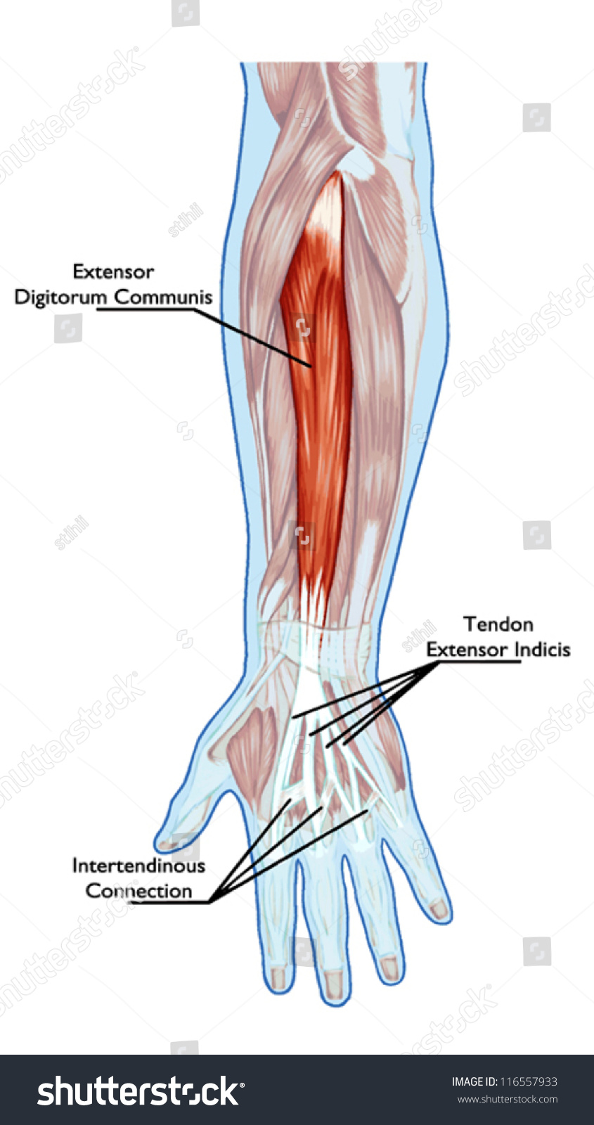
Forearm muscles in the anterior compartment are arranged in superficial, intermediate and deep categories. The superficial group arises mostly from the posterior aspect of the lateral epicondyle of the humerus by a common tendon. Hold your elbow with thumbs up and other 4 extension of index finger. Edc tendons straighten the index, middle, ring and small fingers. A common muscle belly is shared by all the fingers. This does not mean that. Cross sectional anatomy of the upper limb : All 4 muscles have a common origin at the medial epicondyle of the humerus, known as the common flexor tendon. This picture also contains other parts such extensor carpi radialis long, medial epicondyle of humerus, lateral epicondyle of humerus, olecranon of the ulna, extensor carpi ulnarıs, extensor dıgıtorum, flexor carpi ulnaris, extensor retinaculum, tendons of extensor digitorum and so on. Most of these originate from the lateral epicondyle. It turns… inflamed common flexor tendon cft. Important questions on muscles of flexor and extensor compartment of forearm, structures deep and superficial to 3 name the muscles of flexor compartment of forearm supplied by median nerve. Colorful medical illustration of human legs muscles.
Muscles of the forearm that move the wrist, hand, thumb, and digits. Anconeus muscle is a small muscle that is triangular in shape. Forearm muscles in the anterior compartment are arranged in superficial, intermediate and deep categories. A deep layer , intermediate layer and superficial layer. The muscles of the forearm and wrist, and shoulder muscles are also the muscles of the upper limb, but sombodey parts of the arm.
2 Superficial Extensor Muscles And Tendons T Of The Forearm And Download Scientific Diagram from www.researchgate.net They receive additional fibers from the deep fascia of the forearm near the elbow, and from the septa which pass from this fascia between the individual muscles. 3d rendered illustration of the male musculature. It inserted independently into the. By moving the mouse cursor over a particular area of the arm or forearm, this area is highlighted and the labels are displayed: All 4 muscles have a common origin at the medial epicondyle of the humerus, known as the common flexor tendon. Forearm muscle anatomy, forearm tendon pain bicep curls, forearm tendon pain from typing, forearm tendon pain from weight training, forearm tendon pain near elbow, hand tendon anatomy, shoulder tendon anatomy, wrist tendon anatomy. From superior to inferior, origin. A deep layer , intermediate layer and superficial layer.
Cross sectional anatomy of the upper limb :
In the anterior compartment, they are split into three categories: The forearm is the region of the upper limb between the elbow and the wrist. Long flexor tendons extend from the forearm muscles through the wrist and attach to the small bones of the fingers and thumb. Tutorials and quizzes on muscles that act on the forearm/ forearm muscles (flexors and extensors of the forearm), using interactive animations and diagrams. Hold your elbow with thumbs up and other 4 extension of index finger. The superficial group arises mostly from the posterior aspect of the lateral epicondyle of the humerus by a common tendon. When identifying the function of the forearm muscles, it is important to note that any forearm compartment muscle that crosses the elbow joint will act at this joint. See anatomy pictures of the 27 bones in the hand and wrist, how they are connected with tendons and muscles and the nerves that run through the skeletal structure. If you keep your hand flat on a table and. They receive additional fibers from the deep fascia of the forearm near the elbow, and from the septa which pass from this fascia between the individual muscles. The superficial anterior forearm muscles share a common origin on the common flexor tendon that arises from the medial epicondyle of humerus. Anconeus muscle is a small muscle that is triangular in shape. The supinator is a muscle.
12 photos of the forearm tendon anatomy picture. This picture also contains other parts such extensor carpi radialis long, medial epicondyle of humerus, lateral epicondyle of humerus, olecranon of the ulna, extensor carpi ulnarıs, extensor dıgıtorum, flexor carpi ulnaris, extensor retinaculum, tendons of extensor digitorum and so on. Anterior, lateral or posterior compartment. They receive additional fibers from the deep fascia of the forearm near the elbow, and from the septa which pass from this fascia between the individual muscles. Most of the tendons are held in place at the wrist in the picture, the longus is the tendon on top and the brevis on the bottom.
Anatomy Muscular System Hand Forearm Palm Stock Vector Royalty Free 116557933 from image.shutterstock.com All 4 muscles have a common origin at the medial epicondyle of the humerus, known as the common flexor tendon. 3d rendered illustration of the male musculature. The superficial anterior forearm muscles share a common origin on the common flexor tendon that arises from the medial epicondyle of humerus. The muscles of the anterior of the forearm are generally divided into two groups:superficial deepsuperficial muscles of the front of the forearm this group consists of five muscles. It inserted independently into the. All superficial muscles are arises from the medial epicondyle of humerus but they are inserted into the different part except. There are many muscles in the forearm. The anterior forearm muscles are divided into 3 muscular layers ;
While the ventral side of the forearm is not exactly less complicated than the dorsal side, it appears less complicated on the.
The pronator teres has two heads of. Most of these originate from the lateral epicondyle. Important questions on muscles of flexor and extensor compartment of forearm, structures deep and superficial to 3 name the muscles of flexor compartment of forearm supplied by median nerve. The muscles of the forearm are numerous, differ in the variety of functions. Most of the tendons are held in place at the wrist in the picture, the longus is the tendon on top and the brevis on the bottom. The superficial anterior forearm muscles share a common origin on the common flexor tendon that arises from the medial epicondyle of humerus. From superior to inferior, origin. A deep layer , intermediate layer and superficial layer. Muscles of the forearm segregate into these compartments consisting of (1) an anterior group (the flexors seven superficial and five deep muscles occupy the posterior forearm. Anterior, lateral or posterior compartment. The forearm is the region of the upper limb between the elbow and the wrist. 12 (4 superficial + 3 mobile wad + 5 deep). Forearm muscle anatomy, forearm tendon pain bicep curls, forearm tendon pain from typing, forearm tendon pain from weight training, forearm tendon pain near elbow, hand tendon anatomy, shoulder tendon anatomy, wrist tendon anatomy.
When identifying the function of the forearm muscles, it is important to note that any forearm compartment muscle that crosses the elbow joint will act at this joint picture of forearm tendons. The tendons travel down the forearm through a tough band of tissue on top of the wrist.


0 Komentar